

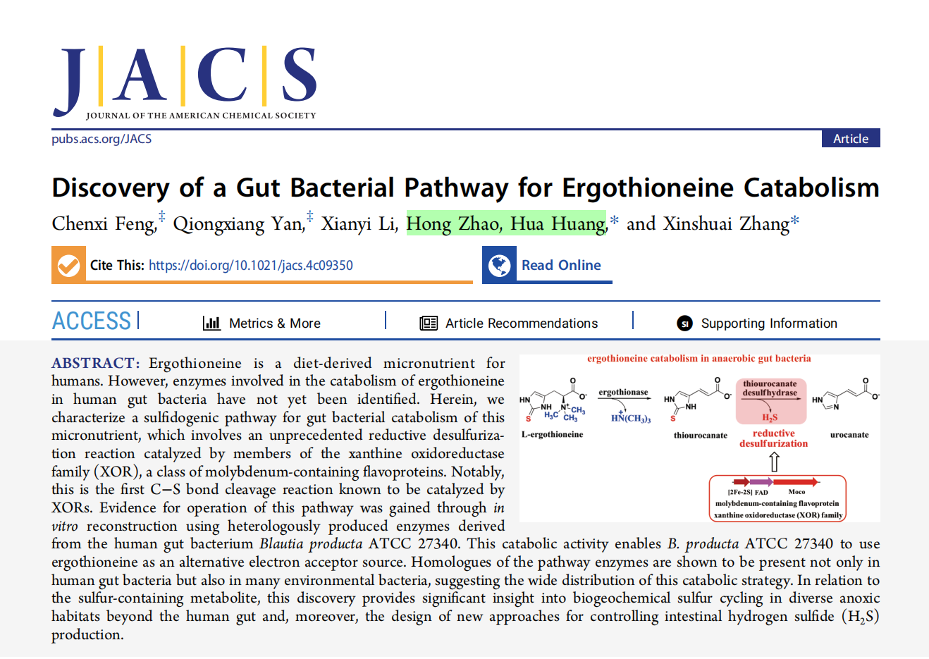
Recently, enzyme product scientists Dr. Hua Huang and Dr. Hong Zhao collaborated with Professor Xinshuai Zhang’s research group at South China Normal University to reveal the gut microbial metabolic pathway of ergothioneine. The relevant research results were recently published in the top academic journal Journal of the American Chemical Society (JACS, impact factor 14.4).
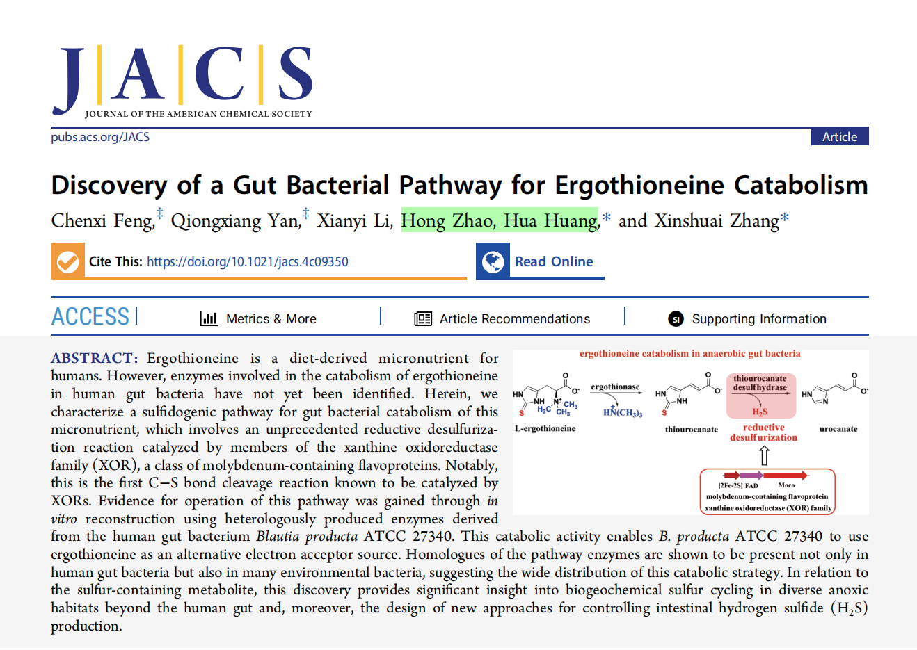
This article reveals the metabolic pathway of ergothioneine via gut microbiota. During metabolism, human gut bacteria ( Blautia producta ATCC 27340) first use an amino acid lyase (PF00221) to cleave ergothioneine trimethylamine to generate thiourocanate. Then, a complex metalloenzyme (Xanthine oxidoreductase, XOR) containing three cofactors—Mo, [2Fe-2S], and FAD—is used to desulfurize it into urocanate (an L -histidine metabolite). This study provides a foundation for research on the mechanism of action of ergothioneine, a common dietary supplement, in the human body.
(Note: JACS is the flagship journal of the American Chemical Society. With its outstanding academic standing and far-reaching influence, it holds a premier position among comprehensive chemistry journals and is hailed as the “holy grail” of chemistry. As the most prestigious journal in the field of chemistry, JACS has become an important reference for measuring the research strength and academic influence of research institutions.)
The following is an analysis of the paper:
Research Project Overview
Ergothioneine is a trimethyl histidine derivative containing a thione structure. It can enter cells and mitochondria through a specific transport protein (SLC22A4) and is one of the few substances with a clear specific transport mechanism [1] . Although mammals cannot directly synthesize ergothioneine, they can absorb and accumulate it from the food chain using specific transport proteins. Some foods, such as mushrooms and black/red beans, contain high concentrations of ergothioneine, and it is also accumulated in many tissues and organs of the human body, especially in organs such as red blood cells, bone marrow, liver and cornea. Experiments have shown that the human body can rapidly absorb exogenously ingested ergothioneine to significantly increase the concentration of ergothioneine in blood and plasma [2] .
Ergothioneine, as a highly effective antioxidant with high safety (6000 times that of Vitamin E ) [3] , has long been recognized as “generally recognized as safe” (GRAS, GRN-000734) by the European Food Safety Authority (EFSA) and the U.S. Food and Drug Administration (FDA), and is now widely used in cosmetic raw materials, dietary supplements, and even food additives [4] . In addition, ergothioneine’s strong reactive oxygen scavenging ability and lipid peroxide inhibition ability also make it a potential [5] . In the field of cosmetics, ergothioneine can absorb ultraviolet rays in a wavelength range similar to DNA, thereby protecting DNA cells in the skin from damage [6] . Therefore, ergothioneine has broad application prospects in various industries such as medicine, food, health products and cosmetics due to its unique biological functions and pharmacological activities. In order to further analyze and explore the various physiological functions and potential applications of ergothioneine, research on the synthetic and catabolic pathways of ergothioneine in organisms is of great significance.
Ergothioneine biosynthesis and metabolism research
The biosynthesis of ergothioneine has been studied in a relatively thorough manner. In its biosynthetic pathway , L -histidine is first methylated by the methyltransferase EgtD (PF10017) to generate L -trimethylhistidine (TMH, Hercynine). Then, in different microbial environments, sulfur addition reactions are carried out using different enzymes and sulfur donors. Therefore, the differences in the biosynthesis of ergothioneine are mainly reflected in the different ways of sulfur addition. To date, three different L -trimethylhistidine sulfation methods have been reported. Under aerobic conditions, aerobic bacteria such as Mycobacterium smegmatis utilize a mononoclear non-heme iron enzyme EgtB (PF12867 & PF03781) to attach the sulfur on the substrate of L -cysteine or γ -glutamyl-cysteine dipeptide ( γ – Glu-Cys, the dipeptide component in glutathione ) to the Hercynine imidazole ring, and finally convert it into ergothioneine (Figure I, Route I) by the lyase EgtE (PF00266). This ergothioneine biosynthesis method mainly exists in aerobic bacteria and fungi [7] . Under anaerobic conditions, the absolutely anaerobic green sulfur bacteria Chlorobium limicola can utilize a thiocyanate generating enzyme EanB (Rhodanese-like The enzyme (PF00581) transfers sulfur, and its sulfur donor is sulfur polymers that exist in the natural environment ( Figure I, Route II). This synthetic pathway is mainly found in anaerobic bacteria and archaea, which also shows that ergothionein is important for life that does not depend on oxygen [8] . In 2022, Seebeck’s group discovered a Mo-dependent ( Metallopterin -dependent) bifunctional (N-terminal tungsten-dependent hydratase and C-terminal cysteine desulfurase) ergothionein synthase MES (PF00384 & PF01568) in the thermophilic anaerobic archaea Caldithrix abyssi. Unlike the EanB enzyme mentioned above, MES uses L -cysteine as a sulfur donor (Figure I, Route III). This ergothionein biosynthesis pathway is also mainly distributed in anaerobic bacteria and archaea [9] .
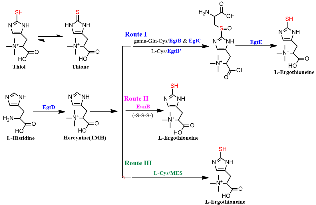
Figure I: Known reported biosynthetic pathways of ergothionein. These include the oxygen-dependent sulfur-producing enzyme pathway (EgtB, Route I) in aerobic bacteria, the thiocyanate-producing enzyme pathway (EanB, Route II) in anaerobic sulfur bacteria, and the Mo-dependent synthase pathway (MES, Route III) in anaerobic thermophilic archaea.
Ergothioneine catabolism study
In recent years, research on ergothioneine catabolism has received increasing attention, and the differential catabolism pathways of ergothioneine in various microorganisms have been revealed. Overall, ergothioneine catabolism mainly revolves around detrimethylamine (TMA) and desulfurization. Ergothioneine is cleaved by the ergothioneine lyase ETL (PF00221, belonging to the same family as L -histidine lyases) to produce trimethylamine and the corresponding uric acid, but there are significant differences in desulfurization; therefore, similar to the biosynthetic diversity of ergothioneine mentioned above, the differences in ergothioneine catabolism are mainly reflected in its different desulfurization mechanisms. In 2020, Professor Shinji Nagata’s research group at Kochi University in Japan discovered a thiourocanic acid desulfurase ETL for the first time, thus initiating a systematic study on the decomposition and metabolism of ergothioneine. This desulfurase belongs to an unknown functional enzyme family DUF917 (PF06032 & PF20906) [10] . The following year, Seebeck’s research group at the University of Basel in Switzerland disclosed for the first time the first complete ergothioneine decomposition pathway involving DUF917 enzyme (Figure II, Route I). This degradation pathway is mainly distributed in soil bacteria (Proteobacteria and Post-myotrophic Bacteria). These soil bacteria do not utilize the antioxidant properties of ergothioneine (direct desulfurization). Their widespread presence reduces the abundance of ergothioneine in the soil in nature, thereby indirectly affecting the absorption of ergothioneine by plants and the overall content of ergothioneine in the food chain [11] .
Previously, Dr. Huang Hua collaborated with Zhang Xinshuai’s research group to discover the oxidative decomposition metabolic pathway of ergothionein in microorganisms using a gene enzyme omics strategy. The relevant results were published in ACS Catalysis in 2022 (impact factor: 12.9). Ergothioneine is converted into ergothioneine sulfinate (Egt-SO₂H) or ergothioneine sulfonate (Egt-SO₃H) after being exposed to oxygen, oxygen free radicals or exerting antioxidant functions. Then , they are desulfurized and desulfurized under the action of metal-dependent ergothioneine sulfinate desulfurase ETSD (SSF51556) and NADPH-dependent ergothioneine sulfinate desulfurase Sulfite lyase (PF02423) (Figure II, Route II, Route III). The ETSD decomposition pathway exists in actinomycetes, which can both synthesize and decompose ergothioneine, indicating their complex sulfur utilization mechanism [12] . Sulfite lyase is widely distributed in soil bacteria such as Proteobacteria, which indirectly indicates that ergothioneine and its sulfonate are widely present in soil [13] .
This study revealed a novel desulfurization mechanism of ergothioneine in human gut microbiota. Members of the gut bacterium *Blautia producta* ATCC 27340, specifically xanthine oxidoreductase (XOR), can perform reductive desulfurization of ergothioneine under anaerobic conditions (Figure II, Route IV). This enzyme is composed of multiple domains containing three cofactors: Mo, [2Fe-2S], and FAD, and exhibits an unknown and complex electron transfer mechanism. This study also provides a foundation for research on the metabolism of ergothioneine in humans.
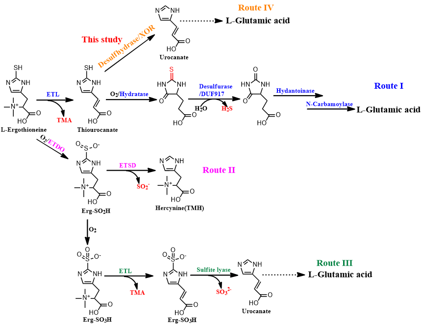
Figure II: Known reported ergothioneine catabolism pathways. Desulfurization metabolism involving DUF917 (Route I), desulfinization metabolism involving ETSD (Route II), desulfonation metabolism involving Sulfite Lyase (Route III), and desulfurization metabolism involving XOR in this study (Route IV).
The above studies on ergothioneine metabolism in nature show that ergothioneine, as a sulfur-containing derivative of L -histidine, has its anabolic and catabolitic metabolisms closely related to L -histidine metabolism, achieving a cycle from L -histidine back to L -histidine metabolism. Meanwhile, compared to the structure of L -histidine, the uniqueness of the ergothioneine molecule mainly lies in the sulfur group on the nitrogen heterocycle; therefore, its metabolic differences revolve around sulfur addition and desulfurization (Figure III).
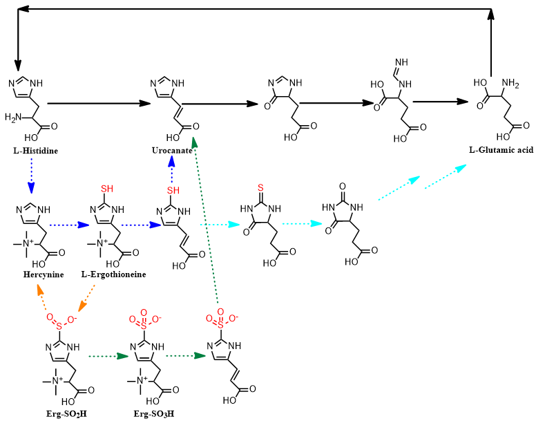
Figure III: Summary of ergothioneine metabolism. Solid lines represent L-histidine metabolism, and dashed lines represent known reported ergothioneine metabolic pathways.
This study is the first to reveal the gut microbial metabolism of ergothioneine.
This thesis continues the experimental “enzymomics” strategy to discover new enzymes and metabolic pathways. Through in-depth analysis of the background differences of the genes corresponding to ergothioneine lyase (ETL, PF00221, currently with 41203 members), it was finally determined that an unknown xanthine oxidoreductase (XOR, Uniprot ID: A0A6P1YXV1, A0A6P1YYW0, A0A6P1YWH0) in the human intestinal bacterium *Blautia producta * ATCC 27340 is involved in new ergothioneine metabolism. This XOR is encoded by three consecutive genes, therefore the enzyme is composed of three subunits: a molybdenum cofactor (Moco) binding domain, an iron-sulfur cluster [2Fe-2S] 2+ binding domain, and a flavin adenine dinucleotide (FAD) binding domain. The known reported function of XOR enzymes is mainly to catalyze the oxidative hydroxylation (oxygenation) of various sp2 hybrid carbons in N-heterocyclic compounds and aldehydes. Through anaerobic expression, purification, and reconstruction tests, the laboratory confirmed that this enzyme possesses the function of desulfurizing thiouric acid (Figure IV). Furthermore, in vivo testing of * B. producta* ATCC 27340 with ergothioneine revealed that this bacterium cannot effectively utilize ergothioneine as a nitrogen or carbon source for cell growth, but primarily uses it as an electron acceptor. When environmental ergothioneine levels are high (concentration above 1 mM), *B. producta * can rapidly absorb external ergothioneine into its cells, demethylate it (TMA), convert it into thiouric acid, and then excrete it. This ergothioneine catabolism mainly occurs within the Firmicutes phylum ( a dominant phylum in the gut microbiota).
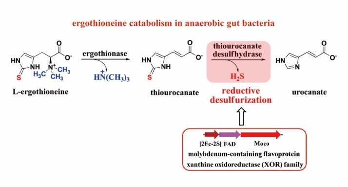
Figure IV: Ergothioneine catabolism pathway involving XOR enzymes in gut microbiota.
Paper link :https://pubs.acs.org/doi/10.1021/jacs.4c09350
References:
[1] Yiwen Han, et al. Crit. Rev. Biotechnol. 2021 , 41 , 580-593;
[2] Irwin K. Cheah, et al. Biochimica Biophysica Acta 201 2 , 1822 , 7 84–793; [3] https://www.businesswire.com/news/home/20110711005157/en/OXIS-International-Top-Anti-Aging-Experts-Worldwide-International
[4] Barry Halliwell, et al. Annu. Rev. Food. Sci. Technol. 2023 , 14 , 323-345;
[5] Irina Borodina, et al. Nutr. Res. Rev. 2020 , 33 , 190-217;
[6] Bindu D. Paul, Antioxid. Redox Signal. 2022 , 36 , 16-18;
[7] Abteilung Physikalische Biochemie, et al. JACS , 2010 , 132 , 6632-6633;
[8] Reto Burn, et al . Angew. Chem. Int. Ed., 2017 , 56 , 12508-12511;
[9] Mariia A. Beliaeva and Florian P. Seebeck, JACS Au 2022 , 2 , 2098−2107;
[10] Hisashi Muramatsu, et al. Biosci. Biotechnol. Biochem. 2021 , 85 , 626-629;
[11] Mariia A. Beliaeva, et al . ACS Chem. Biol. 2021 , 16 , 397−403 ;
[12] Egor Y. Nalivaiko, et al . Angew. Chem. Int. Ed. 2024 , 63 , e20 2318445;
[13] Qiongxiang Yan, et al. ACS Catal. 2022 , 12 , 4825−4832.